Table of Contents >> Show >> Hide
- Stage vs. grade: Two labels, two different jobs
- Symptoms of breast cancer: What to watch for (and why “no symptoms” can still happen)
- How doctors determine breast cancer stage today
- Breast cancer stages, explained in plain English
- Breast cancer grades: What Grade 1, 2, and 3 really mean
- How stage and grade affect treatment decisions
- Outlook and prognosis: What stage and grade can (and can’t) tell you
- Reading your pathology report without spiraling
- Questions to ask your care team (bring this listseriously)
- Experiences: What people often go through with stages and grades (about )
- Conclusion
- SEO tags (JSON)
If you’ve ever felt personally attacked by a pathology report full of letters, numbers, plus signs, and the occasional Roman numeral, you’re not alone.
Breast cancer “stage” and “grade” are two of the most important parts of that alphabet soupbut they’re not the same thing, and mixing them up can make an already stressful moment feel even more confusing.
This guide breaks down the stages and grades of breast cancer in plain English: what they mean, what symptoms can show up (and why early breast cancer can have no symptoms at all),
how doctors figure out the outlook, and what questions help you feel more in control. We’ll keep it accurate, human, and just lightly humorousbecause sometimes you need a laugh and a fact in the same paragraph.
Stage vs. grade: Two labels, two different jobs
Breast cancer stage = where it is and where it’s traveled
Think of stage as the “map.” It describes how much cancer is in the body and how far it has spread. Staging considers:
tumor size, lymph node involvement, and whether cancer has spread to distant organs.
For breast cancer, staging most often uses the TNM system:
T (tumor size/extent), N (nearby lymph nodes), and M (metastasisspread to distant sites).
You’ll also hear about clinical stage (based on exam, imaging, and biopsy before surgery) and pathologic stage (based on what’s found after surgery).
Breast cancer grade = how the cells behave under the microscope
Grade is more like the “personality test” of the cancer cells: how abnormal they look and how quickly they’re likely to grow and spread.
In general, lower-grade cancers tend to grow more slowly than higher-grade cancers.
For many breast cancers, grading uses a system often called the Nottingham (or similar) methodscoring how organized the cells look and how actively they’re dividing.
Grades are usually reported as Grade 1, 2, or 3.
Symptoms of breast cancer: What to watch for (and why “no symptoms” can still happen)
Here’s the tricky truth: early breast cancer may not cause any symptoms, which is one reason screening and routine checkups matter.
Still, many people do notice changes that are worth getting checked.
Common breast cancer symptoms and signs
- A new lump in the breast or underarm
- Thickening or swelling of part of the breast
- Dimpling, puckering, or irritation of breast skin
- Redness, flaking, or scaling of the nipple area or breast skin
- Nipple pulling inward (inversion) or new nipple pain
- Nipple discharge that isn’t breast milk (especially if bloody)
- A change in breast size, shape, or how it feels
- Breast pain that’s new or persistent
When to seek medical care
If you notice a new lump or a change that doesn’t go away, it’s smart to schedule an appointment.
Plenty of breast changes turn out to be non-cancerousbut it’s always better to get clarity than to play “Google roulette” at 2 a.m.
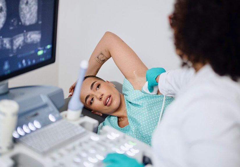
How doctors determine breast cancer stage today
Staging isn’t just one test. It’s a processlike assembling a puzzle where half the pieces come from imaging and the other half come from the lab.
Your care team may use:
- Biopsy (confirms cancer and allows testing of tumor biology)
- Imaging (such as mammogram, ultrasound, MRI; sometimes CT, bone scan, or PET depending on the situation)
- Surgery findings (tumor size, lymph nodes tested, margins)
- Pathology and biomarker tests (ER, PR, HER2, tumor grade, sometimes gene-expression tests)
Why biomarkers matter (and why staging got “smarter”)
Modern breast cancer staging often includes more than anatomy.
Along with TNM, doctors commonly factor in tumor grade, hormone receptor status (estrogen and progesterone receptors),
HER2 status, and sometimes certain gene-expression tests for early-stage cases.
Translation: two tumors that are the same size can behave differentlyand the stage and treatment plan can reflect that.
Breast cancer stages, explained in plain English
Breast cancer stages are typically described as Stage 0 through Stage IV.
Within stages, letters (A, B, sometimes C) add detail. The best way to think about stages is “how far has it spread,” not “how bad is it.”
Stage 0: Non-invasive (carcinoma in situ)
Stage 0 usually refers to ductal carcinoma in situ (DCIS).
In DCIS, abnormal cells are inside a milk duct and have not invaded surrounding breast tissue.
It’s often found on imaging before you’d ever feel a lump.
Stage 0 is highly treatable, but it still needs a real planbecause some cases can progress if left untreated.
Treatment may include surgery (lumpectomy or mastectomy in some situations) and sometimes radiation or medication, depending on risk factors.
Stage I: Small and localized
Stage I generally means an invasive cancer is present, but it’s still small and limited.
Lymph nodes may be negative or show only tiny amounts of cancer in certain scenarios.
A common example: a small tumor found early, removed with surgery, followed by radiation and/or medication based on its biology.
Many stage I cancers have excellent outcomes, especially when treatment is tailored to receptor status and other features.
Stage II: Larger tumor and/or limited lymph node involvement
Stage II often means one of two things: the tumor is bigger, or cancer has spread to a small number of nearby lymph nodes,
usually in the underarm area.
Here’s a helpful mental picture: stage II is still often considered “early-stage” in day-to-day conversation,
but it may require a combination of treatmentssurgery plus radiation, and frequently systemic therapy (like hormone therapy, targeted therapy, and/or chemotherapy).
Stage III: Locally advanced
Stage III is often called locally advanced breast cancer. It typically involves more extensive lymph node involvement
and/or tumor growth into nearby breast structures (for example, skin or chest wall involvement).
Treatment commonly involves a multi-step plan. Many people receive neoadjuvant therapy (treatment before surgery) to shrink the tumor and
make surgery more effective, followed by surgery, radiation, and additional systemic treatment depending on response.
Stage IV: Metastatic breast cancer
Stage IV means the cancer has spread to distant parts of the bodycommonly bone, liver, lung, or brain (though it can spread elsewhere).
Stage IV is also called metastatic breast cancer.
While metastatic breast cancer is usually not considered curable, it is often treatable.
Many people live meaningful lives for years with modern systemic therapies, and treatment choices are strongly guided by biomarkers (ER/PR/HER2) and prior treatments.
Breast cancer grades: What Grade 1, 2, and 3 really mean
If stage is the map, grade is the “how fast is it trying to move?” clue.
Grades are typically:
Grade 1 (low grade / well-differentiated)
Cancer cells look more like normal breast cells and tend to grow and spread more slowly.
Grade 1 often (not always) points to a less aggressive pattern.
Grade 2 (intermediate grade / moderately differentiated)
The middle child of grades: cells look more abnormal than Grade 1 but not as chaotic as Grade 3.
Many breast cancers fall into this category, and treatment decisions rely heavily on other factors too.
Grade 3 (high grade / poorly differentiated)
Cells look very different from normal and tend to grow and spread faster.
Grade 3 doesn’t automatically mean a poor outcome, but it often signals a need for more aggressive systemic therapyespecially depending on the stage and biomarkers.
How stage and grade affect treatment decisions
Breast cancer treatment isn’t one-size-fits-all. Most plans are built from a few “main categories,” combined based on stage, grade, and tumor biology.
Local treatments (focused on the breast/nearby area)
- Surgery (lumpectomy or mastectomy; lymph node evaluation may include sentinel node biopsy)
- Radiation therapy (often after lumpectomy; sometimes after mastectomy depending on nodes and tumor features)
Systemic treatments (travel through the body)
- Hormone therapy (for ER/PR-positive cancers)
- HER2-targeted therapy (for HER2-positive cancers)
- Chemotherapy (more common with higher stage, higher grade, or higher-risk biology)
- Immunotherapy (used in specific situations, such as certain triple-negative breast cancers)
- Other targeted therapies (based on biomarkers and prior treatments)
A quick example (not a diagnosisjust a demo)
Imagine two people both have a 2 cm tumor and a negative lymph node biopsy. That might sound similar at first.
But if one tumor is Grade 1 and hormone receptor-positive, and the other is Grade 3 and triple-negative,
the recommended systemic therapy can look very differenteven if the anatomic stage is similar.
Stage tells you where; grade and biomarkers help predict behavior and guide the smartest treatment mix.
Outlook and prognosis: What stage and grade can (and can’t) tell you
It’s normal to jump straight to “What’s my outlook?” but prognosis is not a single number.
Doctors estimate prognosis by looking at the full picture: stage, grade, ER/PR/HER2 status, tumor type, response to treatment, and overall health.
Survival rates: helpful for context, not fortune-telling
Large population statistics can provide perspective, but they can’t predict an individual outcomeespecially because treatments keep improving.
In the U.S., five-year relative survival rates for invasive breast cancer are highest when cancer is localized and lower when it has spread regionally or distantly.
- Localized (confined to the breast): typically around the high 90% range
- Regional (nearby lymph nodes/structures): typically in the mid-to-high 80% range
- Distant (metastatic): typically around the low 30% range
Biomarkers matter too. For example, hormone receptor and HER2 status can influence both treatment response and survival patterns.
That’s why you may hear your team talk about a “prognostic stage” that blends anatomic findings with biology.
What can improve outlook?
- Earlier detection (many early cancers are treated very successfully)
- Tailored therapy based on ER/PR/HER2 and other tests
- Good response to treatmentespecially neoadjuvant therapy when used
- Finishing the full treatment plan, including long-term therapies when prescribed
- Support and follow-up (side effects and mental health matter; you’re not a robot)
Reading your pathology report without spiraling
The pathology report can feel like it was written by someone who charges by the acronym.
These are common items you’ll see and what they generally mean:
- Tumor type (such as invasive ductal carcinoma, DCIS, inflammatory breast cancer)
- Tumor size (part of the “T” in TNM)
- Grade (1–3)
- Margins (whether cancer cells are at the edge of removed tissue)
- Lymph node results (part of the “N” in TNM)
- ER/PR status (hormone receptors)
- HER2 status (HER2 protein/amplification)
- Sometimes gene-expression tests (in specific early-stage scenarios)
If you don’t understand a term, ask for a plain-language explanation. You deserve one.
A helpful question is: “Can you explain how my stage, grade, and biomarkers work together to guide treatment?”
Questions to ask your care team (bring this listseriously)
- What is my clinical stage right now? Will it change after surgery?
- What is my tumor grade, and what does it suggest about growth rate?
- What are my ER, PR, and HER2 results?
- Am I being staged using an anatomic approach, a prognostic approach, or both?
- Do I need genetic counseling or testing based on my age/family history?
- Is treatment recommended before surgery (neoadjuvant), and why?
- What treatments are meant to reduce recurrence risk?
- What side effects should I expectand what can we do to manage them early?
- What follow-up schedule will I have after treatment?
Experiences: What people often go through with stages and grades (about )
Facts are essential, but so is the human side. When people talk about their experiences with breast cancer stages and grades, a few themes come up again and againregardless of age, background, or exact diagnosis.
First, there’s the waiting. Waiting for biopsy results. Waiting for imaging. Waiting for the phone call that changes your week (or your life). Many people say the time between “something looks suspicious” and “here’s the full stage and plan” is the most emotionally intense part, because your imagination fills every quiet moment. It’s common to feel fine one minute and overwhelmed the next, especially when you’re trying to keep everyday life goingschool, work, family, meals, and all the normal stuff that doesn’t pause just because your body decided to be dramatic.
Then comes the terminology shock. People often describe seeing “Stage II” or “Grade 3” and feeling like those labels are a verdict, not a description. In real life, your care team is using them as organizing toolsways to match you to the most effective treatmentsbut emotionally, it can land like a stamp on your forehead. A surprisingly helpful experience for many patients is having a clinician draw a simple chart: stage on one side (where it is), grade on the other (how it looks/behaves), and biomarkers underneath (what it responds to). Once those pieces are separated, the whole situation often feels more navigable.
People also talk about “control points.” You can’t control a diagnosis, but you can control the next step: showing up to appointments, writing questions down, bringing a trusted person (or asking to record the conversation if allowed), and requesting a second opinion when you need reassurance. Many patients say the moment they started keeping a small notebookmedication names, appointment dates, “what does this mean?” questionswas the moment anxiety stopped running the entire show.
Another common experience is the emotional whiplash of treatment planning. A stage 0 or stage I diagnosis can still involve major decisions about surgery and radiation. A stage III diagnosis can come with months of treatment that feel like a second job. People frequently say the hardest part isn’t just the medical logisticsit’s the identity shift: realizing you’re now someone who has to think about receptors, side effects, and follow-up scans. Many find comfort in support groups (online or in-person), therapy, faith communities, or just a tight circle of friends who can handle both serious conversations and normal ones.
Finally, there’s the long view. Whether someone finishes treatment and moves into follow-up care, or lives with metastatic breast cancer and ongoing therapy, many describe learning a new rhythm: celebrating stable scans, adjusting to medication routines, and recognizing that fear can exist alongside hope. If there’s a takeaway from these shared experiences, it’s this: understanding your stage and grade doesn’t just inform treatmentit can also replace some fear with clarity, and clarity is a kind of relief.
Conclusion
The stages and grades of breast cancer are powerful tools for understanding a diagnosis, not a summary of who you are or what’s possible.
Stage describes how far cancer has spread. Grade describes how the cancer cells look and how quickly they may grow. Add in biomarkers like ER, PR, and HER2,
and your care team can build a treatment plan that’s more personalized than ever.
If you’re facing a diagnosisyour own or someone you loveask questions, take notes, and remember: you’re allowed to request explanations in plain English.
The goal isn’t just to treat cancer. The goal is to help you live well through the process, with the best evidence guiding every step.